Skip to content Skip to sidebar Skip to footer

Ct Scan Block Diagram

Ct Scanner Block Diagram Working Operation Construction Applications D E Notes

Ct Scanner Block Diagram Working Operation Construction Applications D E Notes

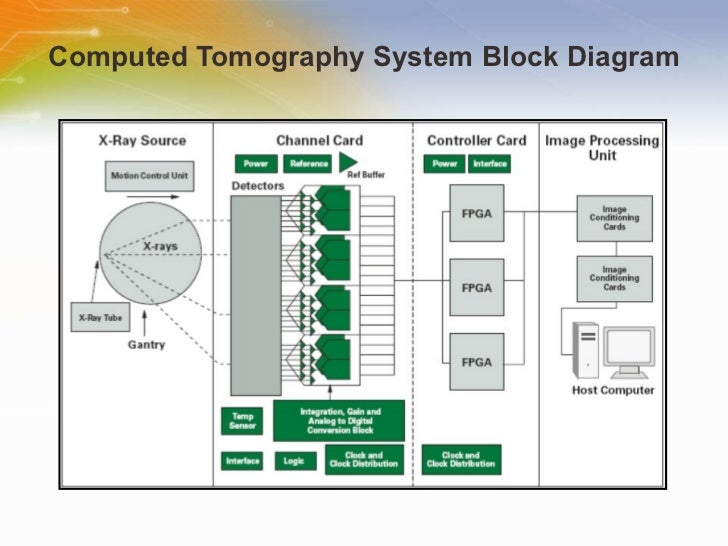
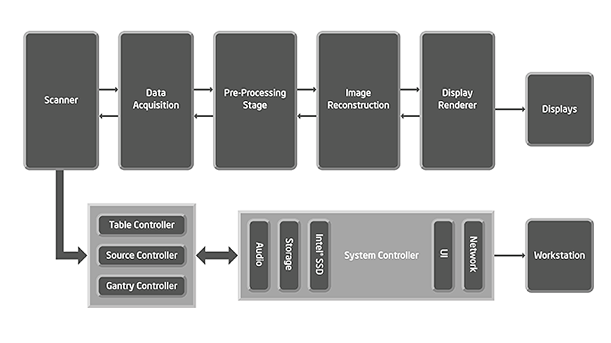
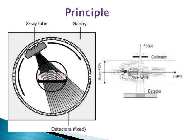
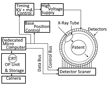
Ct scan block diagram. MRI System Block Diagram RF amp spectrometer rfcoil gradient coil X amp Y amp Z amp 2. Computed tomography CT produces 3D images of internal human body parts from a large series of two-dimensional X-ray images taken around a single axis of rotation. Block Diagram of CT Scanner Operations of CT Scanner High voltage supply drives the x-ray tube that can be mechanically rotated along the circumference of a gantry.

CT scans produce 2-dimensional images of a slice or section of the body but the data can also be used to construct 3-dimensional images. Your message goes here. History 1924 - mathematical theory of thomographic image.

Each picture also called a slice shows a few layers of your. 32-3 CT scan of the Figure 32-2 circuit board from System A showing solder locations 19 32-4 CT scan of the Figure 32-2 circuit board from System A showing the internal structure of the relays 20 32-5 Printed circuit board test phantom 20 32-6 CT scan of the. Very-high-speed computers are necessary to process the images collected in this manner.

A CT scan can be compared to. When compared with a traditional X-ray radiography a CT image exhibits significantly improved contrast. A CT scan or computed tomography scan formerly known as computed axial tomography or CAT scan is a medical imaging technique used in radiology to get detailed images of the body noninvasively for diagnostic purposes.

CT Scanner design resources and block diagram. CT scan tubes often contain more than one size of focal spot. 05 and 10 mm are the common size of focal spot.

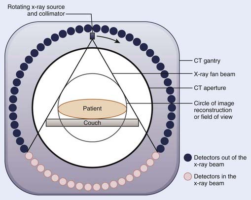
In a spiral-CT system the patient is slowly moved into the center of the gantry while the x-ray source and detector rotate about the patient. 12 hours ago Delete Reply Block. Just like as in standard xray tubes because of reduced penumbra small focal spot in Computed tomography tubes produce sharper images like better spatial resolution but because they concentrate heat onto smaller area of the anode.

A b Diagram a and axial multidetector CT image b show the antecrural technique for celiac plexus block in which the neurolytic solution spreads anterolateral to the aorta causing targeted destruction of the celiac ganglia arrow in b. CT scanners use a rotating x-ray tube and a row of detectors placed in the gantry to.

Ct Scan In Hindi Ct Scan Working Principal Youtube

Ct Scan In Hindi Ct Scan Working Principal Youtube

Ct Scan Block Diagram Explanation Ct Scan Machine

Ct Scan Block Diagram Explanation Ct Scan Machine

Computerized Axial Tomography How It Works Electronics And Communication Study Materials

Computerized Axial Tomography How It Works Electronics And Communication Study Materials

Equipment For Ct Book Chapter Iopscience

Equipment For Ct Book Chapter Iopscience

Block Scheme Of Scanned Electron Beam X Ray Ct Scanner Download Scientific Diagram

Block Scheme Of Scanned Electron Beam X Ray Ct Scanner Download Scientific Diagram

Ct Scan Block Diagram Ct Scan Machine

Ct Scan Block Diagram Ct Scan Machine

Ct Scanner Scanner Block Diagram Diagram

Ct Scanner Scanner Block Diagram Diagram

Block Diagram Of The Experimental Setup The Needle Pose Is Measured Download Scientific Diagram

Block Diagram Of The Experimental Setup The Needle Pose Is Measured Download Scientific Diagram

Electromagnetic Tomography For Medical And Industrial Applications Challenges And Opportunities Ieee Life Sciences

Electromagnetic Tomography For Medical And Industrial Applications Challenges And Opportunities Ieee Life Sciences

Figure 3 From Physiological Gating Of The Mars Spectral Micro Ct Scanner Semantic Scholar

Figure 3 From Physiological Gating Of The Mars Spectral Micro Ct Scanner Semantic Scholar

A Block Diagram Showing The Connections Required To Integrate The Download Scientific Diagram

A Block Diagram Showing The Connections Required To Integrate The Download Scientific Diagram

Gt 5948 Diagram Of Ct Scanner Download Diagram

Gt 5948 Diagram Of Ct Scanner Download Diagram

Figure 2 From Three Dimensional Imaging Of Teeth And Jaw Bones Using Fluoroscopic Computed Tomography Fct Semantic Scholar

Figure 2 From Three Dimensional Imaging Of Teeth And Jaw Bones Using Fluoroscopic Computed Tomography Fct Semantic Scholar

Ct Scanner Solution

Ct Scanner Solution

Ai Ct Scan Analysis For Covid 19 Detection And Patient Monitoring Synced

Ai Ct Scan Analysis For Covid 19 Detection And Patient Monitoring Synced

High End Medical Imaging

High End Medical Imaging

Gt 5948 Diagram Of Ct Scanner Download Diagram

Gt 5948 Diagram Of Ct Scanner Download Diagram

Computerized Tomography An Overview Sciencedirect Topics

Computerized Tomography An Overview Sciencedirect Topics

Medical Image Processing From Formation To Interpretation Analog Devices

Medical Image Processing From Formation To Interpretation Analog Devices

Effective And Reliable Framework For Lung Nodules Detection From Ct Scan Images Abstract Europe Pmc

Effective And Reliable Framework For Lung Nodules Detection From Ct Scan Images Abstract Europe Pmc

X Rays Definition Block Diagram And Working Of X Ray Machine Radiology Schools X Ray Radiology Student

X Rays Definition Block Diagram And Working Of X Ray Machine Radiology Schools X Ray Radiology Student

Block Scheme Of Scanned Electron Beam X Ray Ct Scanner Download Scientific Diagram

Block Scheme Of Scanned Electron Beam X Ray Ct Scanner Download Scientific Diagram

What Are Ct Scans And How Do They Work Omega Pds

What Are Ct Scans And How Do They Work Omega Pds

1

1

Immobilization And Simulation Clinical Gate

Immobilization And Simulation Clinical Gate

How A Micro Ct Scanner Works Micro Photonics

How A Micro Ct Scanner Works Micro Photonics

Magnetic Resonance Imaging Wikipedia

Magnetic Resonance Imaging Wikipedia

Covid 19 Pneumonia Infection Control Protocol Inside Computed Tomography Suites Springerlink

Covid 19 Pneumonia Infection Control Protocol Inside Computed Tomography Suites Springerlink

Ct Scan

Ct Scan

2

2

Bridge Circuit Under Ac Dc Dc Dc Circuits 11909 Next Gr

Bridge Circuit Under Ac Dc Dc Dc Circuits 11909 Next Gr

Ct Imaging By Elizabeth Swanson Openwetware

Ct Imaging By Elizabeth Swanson Openwetware

Figure 16 State Of The Art Of Ct Detectors And Sources A Literature Review Springerlink

Figure 16 State Of The Art Of Ct Detectors And Sources A Literature Review Springerlink

Entropy Free Full Text Classification Of Covid 19 Coronavirus Pneumonia And Healthy Lungs In Ct Scans Using Q Deformed Entropy And Deep Learning Features Html

Entropy Free Full Text Classification Of Covid 19 Coronavirus Pneumonia And Healthy Lungs In Ct Scans Using Q Deformed Entropy And Deep Learning Features Html

2

2

Ct Block Diagram Computer Engineering Personal Computers

Ct Block Diagram Computer Engineering Personal Computers

Ct Scan And It S Block Diagram Youtube

Ct Scan And It S Block Diagram Youtube

Identifying Covid19 From Chest Ct Images A Deep Convolutional Neural Networks Based Approach

Identifying Covid19 From Chest Ct Images A Deep Convolutional Neural Networks Based Approach

Gt 5948 Diagram Of Ct Scanner Download Diagram

Gt 5948 Diagram Of Ct Scanner Download Diagram

A Frameless Armless Navigational System For Computer Assisted Neurosurgery In Journal Of Neurosurgery Volume 74 Issue 5 1991

A Frameless Armless Navigational System For Computer Assisted Neurosurgery In Journal Of Neurosurgery Volume 74 Issue 5 1991

Block Diagram Showing The Image Analysis Steps For A Preoperative Download Scientific Diagram

Block Diagram Showing The Image Analysis Steps For A Preoperative Download Scientific Diagram

Computer Tomography By Moustafa M Mohamed Introduction To Medical Imaging Uses Of Medical Imaging Obtain Information About Internal Body Organs Or The Ppt Download

Computer Tomography By Moustafa M Mohamed Introduction To Medical Imaging Uses Of Medical Imaging Obtain Information About Internal Body Organs Or The Ppt Download

Region Of Interest Based Medical Image Compression Salih Burak Gokturk Abstract Ct Or Mri Medical Imaging Produces Digital Form Of Human Body Pictures There Exists A Need For Compression Of These Images For Storage And Communication Purposes

Region Of Interest Based Medical Image Compression Salih Burak Gokturk Abstract Ct Or Mri Medical Imaging Produces Digital Form Of Human Body Pictures There Exists A Need For Compression Of These Images For Storage And Communication Purposes

2

2

2

2

Current Trends In Pet And Combined Pet Ct And Pet Mr Systems Design Sciencedirect

Current Trends In Pet And Combined Pet Ct And Pet Mr Systems Design Sciencedirect

Breaking Through The Speed Barrier Advancements In High Speed Imaging Intechopen

Breaking Through The Speed Barrier Advancements In High Speed Imaging Intechopen

Pet Scanner Diagram Page 3 Line 17qq Com

Pet Scanner Diagram Page 3 Line 17qq Com

1

1

Copy of CT Scan --You can edit this template and create your own diagramCreately diagrams can be exported and added to Word PPT powerpoint Excel Visio or any other document.

Computed tomography CT produces 3D images of internal human body parts from a large series of two-dimensional X-ray images taken around a single axis of rotation. Please click a block in the diagram to the left to find products. Please click a block in the diagram to the left to find products. 12 hours ago Delete Reply Block. A b Diagram a and axial multidetector CT image b show the antecrural technique for celiac plexus block in which the neurolytic solution spreads anterolateral to the aorta causing targeted destruction of the celiac ganglia arrow in b. Computed tomography CT produces 3D images of internal human body parts from a large series of two-dimensional X-ray images taken around a single axis of rotation. CT scan tubes often contain more than one size of focal spot. 32-3 CT scan of the Figure 32-2 circuit board from System A showing solder locations 19 32-4 CT scan of the Figure 32-2 circuit board from System A showing the internal structure of the relays 20 32-5 Printed circuit board test phantom 20 32-6 CT scan of the. 1971 - first commercial CT Sir Godfrey Hounsfield 1974 - first 3rd generation CT 1979 - Nobel price Cormack Hounsfield 1989 - single-row CT 1994 - double-row spiral CT 2001 - 16-row spiral CT 2007 - 320-row spiral CT.


Each picture also called a slice shows a few layers of your. A CT scan can be compared to. 05 and 10 mm are the common size of focal spot. A b Diagram a and axial multidetector CT image b show the antecrural technique for celiac plexus block in which the neurolytic solution spreads anterolateral to the aorta causing targeted destruction of the celiac ganglia arrow in b. Just like as in standard xray tubes because of reduced penumbra small focal spot in Computed tomography tubes produce sharper images like better spatial resolution but because they concentrate heat onto smaller area of the anode. When compared with a traditional X-ray radiography a CT image exhibits significantly improved contrast. Sophisticated tomographic imaging techniques are used to produce the required image.
Post a Comment for "Ct Scan Block Diagram"